Background: Castleman disease (CD) is a rare, non-clonal lymphoproliferative disorder that is uncommon in children and classified as unicentric or multicentric, with multicentric disease further divided by HHV-8/HIV status into HHV-8-associated and idiopathic (HHV-8-negative) forms. Gastrointestinal, and specifically gastric, involvement is exceptionally rare in any age group and has been described almost exclusively as unicentric disease. Acute gastric haemorrhage as a presenting feature of multicentric CD in a young child appears unreported.
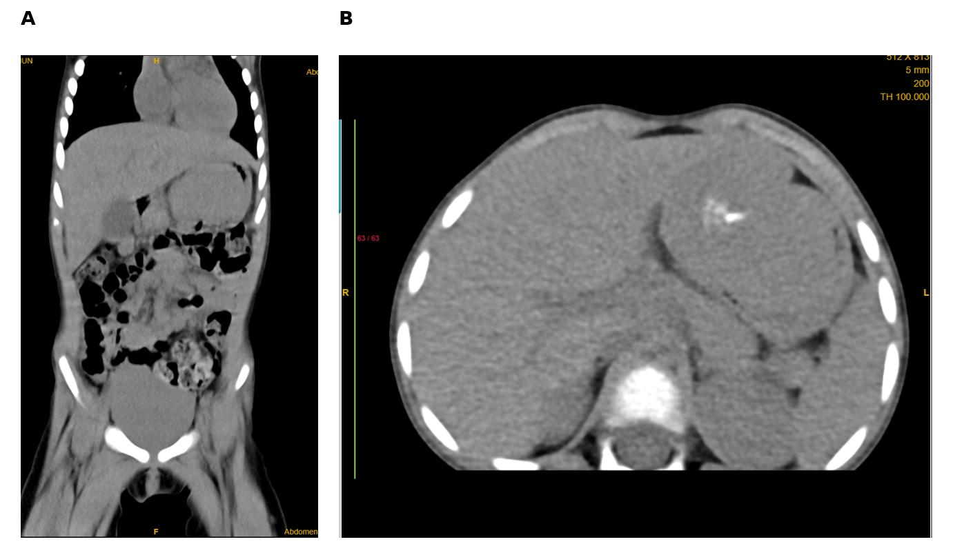
Case Presentation: A previously well 2-year-old girl presented with a one-week history of abdominal pain followed by three episodes of frank haematemesis. Bloods revealed iron deficiency anaemia, mildly elevated inflammatory markers and IL-6, with negative HIV and HHV-8 serology. Ultrasound and contrast-enhanced CT demonstrated a large enhancing gastric mass in the left epigastrium (Figure 1). Endoscopic biopsy was non-diagnostic, and given the high risk of recurrent bleeding, she underwent open partial gastrectomy with excision of prominent pyloric and coeliac nodes. Histopathology initially suggested an inflammatory myofibroblastic tumour; however, immunohistochemistry (CD20+, CD3+, CD138+, CD21/CD23+, polytypic kappa/lambda, ALK-negative, HHV-8 LANA-1-negative) confirmed the plasma-cell/mixed variant of Castleman disease. Staging CT demonstrated multi-station lymphadenopathy, confirming multicentric, HHV-8-negative disease. She was treated with corticosteroids followed by tocilizumab, with resolution of symptoms, normalisation of inflammatory markers, and radiological regression of lymphadenopathy, remaining in clinical and radiological remission at 12 months of follow-up.
Learning Points: To our knowledge, this is the first reported paediatric case of multicentric Castleman disease presenting with acute gastric haemorrhage. This case highlights three key learning points. First, Castleman disease should be included in the differential diagnosis of an acutely bleeding gastric mass in young children, alongside gastrointestinal stromal tumour and inflammatory myofibroblastic tumour, even in the absence of classical systemic features of CD such as fever or chronic diarrhoea. Second, initial histopathology can closely mimic inflammatory myofibroblastic tumour, and a full immunohistochemical panel — including ALK and HHV-8 LANA-1 staining — is essential to reach the correct diagnosis and avoid mislabelling. Third, combined surgical resection of the mass-forming lesion followed by anti-IL-6-directed therapy (tocilizumab) achieved sustained remission in this case, supporting this approach for multicentric paediatric CD presenting acutely with a resectable mass. Clinicians managing young children with unexplained gastric masses and gastrointestinal bleeding should consider CD in their differential and pursue immunohistochemical confirmation, since the distinction carries direct management implications.