Background: Periorbital infections in young infants are uncommon but potentially serious, risking progression to orbital cellulitis, abscess formation, and vision-threatening complications. Presentation may be subtle in neonates, making red flags for deeper tissue involvement essential to recognize. We report a case of periorbital abscess in a 1-month-old infant, highlighting the diagnostic and management challenges in this age group.
Case Presentation: A 1-month-6-day-old male infant presented with a 3-day history of continuous fever and left eye swelling, initially assessed as a left palpebral infection, having received topical ointment and oral amoxicillin without improvement. Trauma/laceration and insect bite were denied; maternal peripartum Staphylococcus colonization status was not available. On admission, he was febrile (40°C) with progressively worsening periorbital edema and erythema; systemic examination was otherwise unremarkable, and he remained hemodynamically stable. Initial labs showed elevated infection markers.
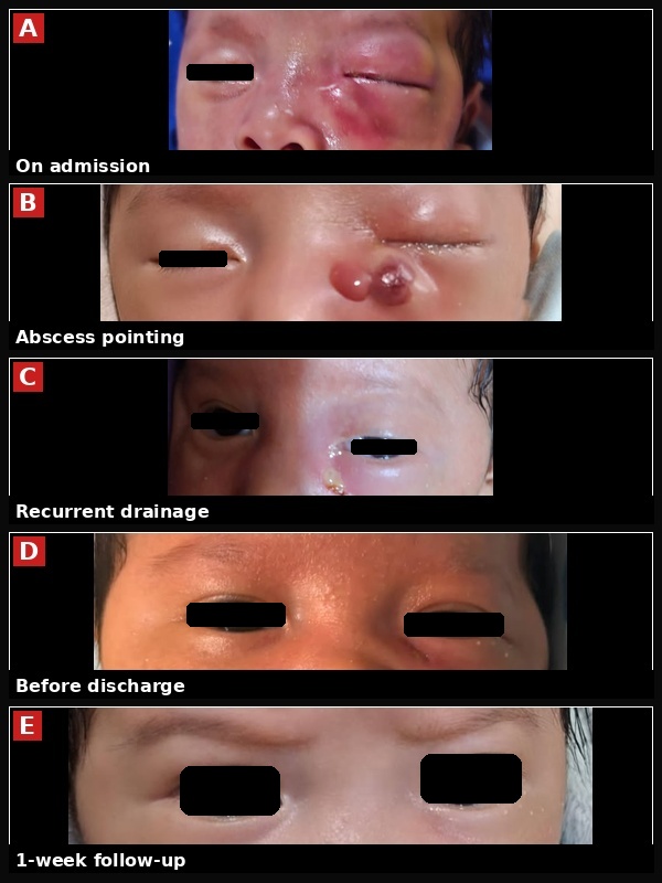
Intravenous ceftriaxone and metronidazole were started; paediatric surgery was consulted but did not advise intervention. Over the first 4 days, fever and swelling improved, but on day 5 swelling recurred afebrile with copious purulent discharge; repeat blood count showed worsening infection markers, prompting a change to meropenem. Ophthalmology was consulted, topical therapy adjusted, and contrast-enhanced orbital computed tomography (CT) obtained, showing a hypodense, rim-enhancing collection (23 Hounsfield units) in the left infero-medial orbit (2.2 × 0.9 × 1.2 cm) with mass effect on the medial and inferior rectus muscles, without intracranial extension (differential: infection vs. mass); meropenem was continued. After 3 further days, swelling and purulent discharge recurred, with repeat markers still unimproved, prompting a change to ampicillin-sulbactam, with clindamycin added on ophthalmology's recommendation. Surgical drainage was deferred given the recurrent spontaneous discharge. Intravenous antibiotics were continued for 14 days total, with clinical and laboratory improvement; at discharge and 1-week follow-up he was clinically well with resolved swelling and normalized infection markers, though mild anaemia persisted (Figure 1). Pus culture and repeat imaging were not obtained, due to resource constraints.
Learning Points: This case illustrates that periorbital abscess, though rare in early infancy, should be considered when swelling fails oral or topical therapy, or progresses despite parenteral treatment. Serial monitoring of inflammatory markers proved essential, prompting sequential escalation to meropenem, then ampicillin-sulbactam with clindamycin. Imaging helped confirm the diagnosis, though the CT differential of infection vs. mass shows imaging needs clinical correlation; here, spontaneous drainage allowed a non-surgical approach to succeed despite repeated multidisciplinary reassessment. A limitation is the absence of microbiological confirmation and repeat imaging, due to resource constraints. This means a resistant organism such as methicillin-resistant Staphylococcus aureus — a recognized pathogen in paediatric periorbital/orbital abscesses — could not be excluded, and the repeated escalation, including a carbapenem, may reflect this uncertainty, reinforcing the value of cultures when feasible. Recurrent or severe abscess-forming infections in infancy also warrant considering an underlying immunodeficiency, though one responsive episode does not warrant work-up. This case underscores the value of vigilant reassessment, judicious antibiotic escalation guided by clinical and laboratory trends, and a pragmatic, resource-conscious multidisciplinary approach.