Objective: Deep cerebral venous thrombosis (DCVT) involves the internal cerebral veins and often co-exists with dural venous thrombosis. Although presentations vary, DCVT often manifests with a more acute and severe trajectory. Typical imaging findings include T2 hyperintense swelling of the thalami and basal ganglia, with or without haemorrhage. We present a child with extensive bilateral DCVT initially treated as acute necrotising encephalitis (ANE).
Method: A retrospective analysis of case notes.
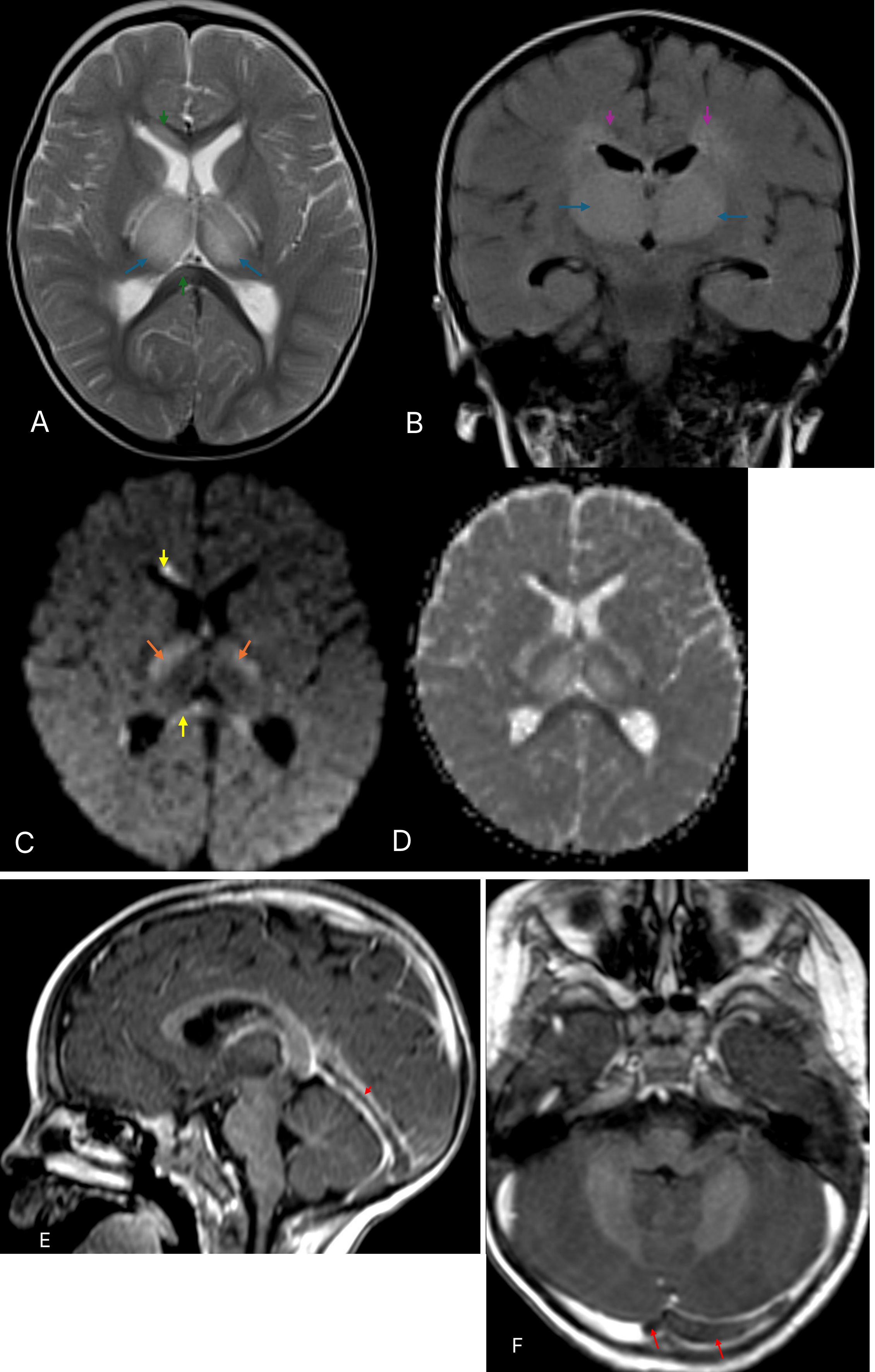
Results: A previously well, developmentally normal,15-month-old boy presented with a 3-day history of fever, diarrhoea, and vomiting followed by rapid-onset encephalopathy. On examination, he was irritable with a fluctuating GCS. There was generalised hypotonia, brisk lower limb reflexes, and intermittent stiffening of his legs on crying, but no clear evidence of seizure activity. There was a significant microcytic anaemia but normal transaminases and inflammatory markers, and CSF was unrevealing. Respiratory PCR was positive for Rhinovirus. An initial CT head demonstrated hypodense thalami and basal ganglia. Intravenous methylprednisolone was commenced for suspected ANE. MR imaging demonstrated significant bilateral thalamic swelling with florid T2/FLAIR hyperintensities and associated diffusion restriction, with additional signal changes involving the corpus callosum, corona radiata and posterior limbs of the internal capsules (PLIC), but no haemorrhage (Figure 1). Dural venous sinus thrombosis involving the straight sinus, vein of Galen and transverse sinuses was identified on MR imaging and subsequently confirmed on CT venography.
He was commenced on anticoagulation and treatment for iron deficiency anaemia and made a full neurological recovery within 2 weeks.
Conclusion: Iron deficiency anaemia is a known risk factor for cerebral venous thrombosis. DCVT is a recognised mimic of ANE in children with several cases having been described in literature, highlighting the diagnostic challenges associated with overlapping clinical and radiological features. Early differentiation with venography should be considered, especially in atypical cases, to support prompt diagnosis and initiation of anticoagulant therapy.