Background: Bronchiolitis is a common lower respiratory tract infection most commonly due to RSV with peak incidence in infants less than a year old, 1.5-2 times more common in boys than girls. It is typically self-limiting and requires solely supportive management, however, in some cases, acute complications such as bacterial infection, respiratory distress, hypoxia and apnoea may occur, requiring admission or medical management. Sequelae can also arise such as recurrent wheeze and asthma. Less experienced or informed health professionals may be tempted to investigate and manage like adults, and this can lead to the over-use of chest x-rays in children and avoidable exposure to radiation. The National Institute of Clinical Excellence (NICE) in England have published guidelines in bronchiolitis management however adherence is lacking. “Do not routinely perform a chest x-ray in children with bronchiolitis.” - NICE.
Methods: Retrospective clinical audit of infants aged 0-24 months who presented to Paediatric Emergency Department at Royal Albert Edward Infirmary A&E between January 2026 and April 2026 in first presentation of illness with a clinical diagnosis of bronchiolitis were included. 130 out of 171 met the criteria. Exclusion criteria included those with supplemental oxygen requirements, severe respiratory distress, significant comorbidity such as chronic lung disease, immunodeficiency, neuromuscular disorders, congenital heart disease and previous CXR for the same illness within 2 weeks. Assessing those who received CXR and why, who reviewed, whether they were admitted and clinical uncertainty.
Results: 16 out of 130 (12.3%) of patients received chest x-rays, 12 out of 130 (9.2%) were admitted. Of those who received chest x-rays,11 out of 16 (68.8%) had no justifiable reason as per criteria set out by NICE guidelines. There was diagnostic uncertainty in 20% of cases. First assessor level was resident doctor in 81.4% of cases. Of the chest x-rays performed, 75% were reported normal. 81.8% of unjustified chest x-rays were reported as normal, meanwhile 60% of justified CXR’s were normal. Two abnormalities noted within the unjustified category: 1. Bilateral mild peri bronchial wall thickening. Otherwise, No focal lung lesions. 2. Small ill-defined right paracardial patchy opacity could suggest early infective process. Otherwise, no focal lung lesions.
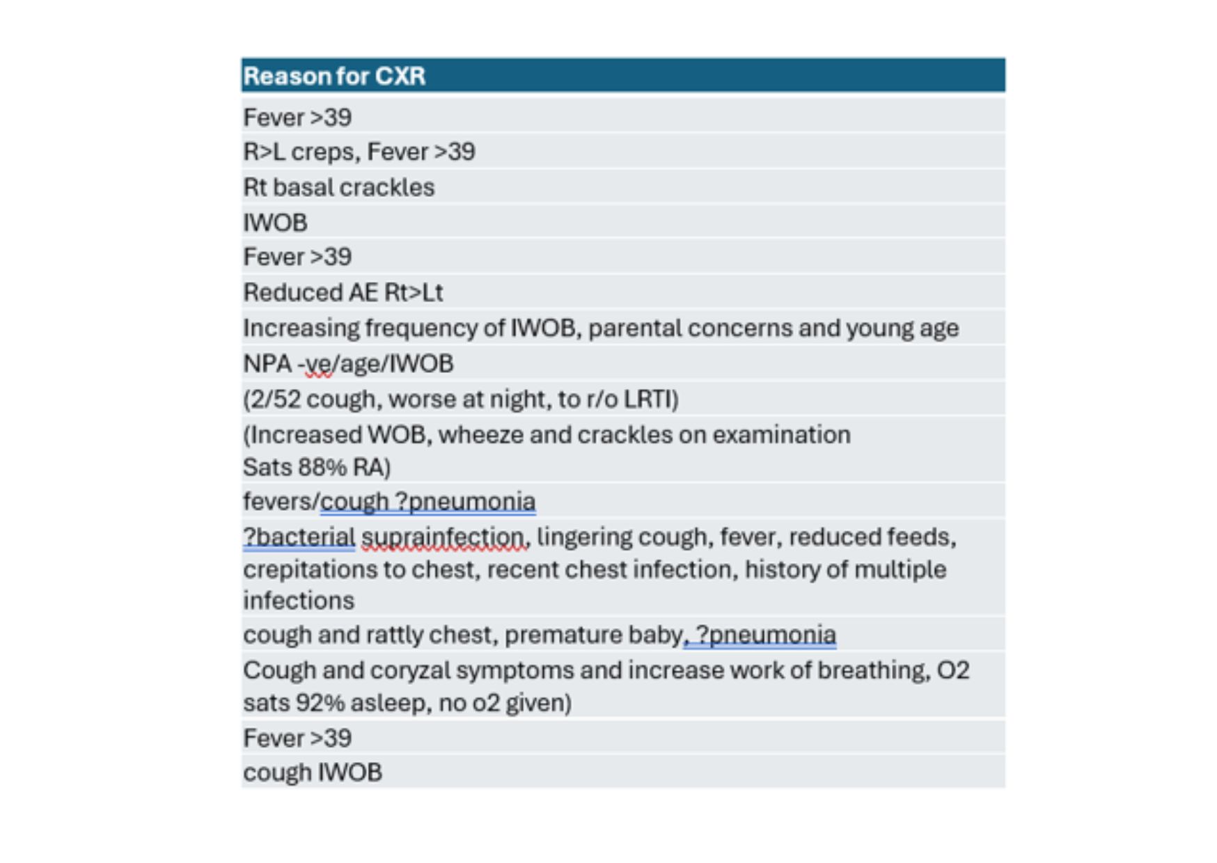
Two abnormalities noted within the justified category: 1. Right retrocardiac and Right lower zonal increased opacification silhouetting Right hemidiaphragm likely infective. 2. Mild peri-bronchial cuffing suggesting small airways inflammation. There was a strong positive correlation between those who received CXR and those who were admitted φ = 0.528, p=0.0000031. A statistically significant moderate correlation existed between those who received a CXR and diagnostic uncertainty p=0.0006. While first assessor was commonly resident, there was no statistical correlation (p=0.1915). See Reason for CXR table (Table 1).
Conclusions: 87.7% did not receive chest x-rays. Which reflects sound clinical judgment in the majority of cases, but this is below the 100% target. Some potential reasons may be clinical uncertainty, in which case a CXR can help differentiate. This audit revealed level of confidence was more likely the reason for CXR request than assessor level.