Background: Transient Neonatal Pustular Melanosis (TNPM) is a benign, self-limited vesiculopustular eruption of the newborn. Small pustules on a non-erythematous base are usually present at birth or appear within the first day of life. They usually rupture spontaneously within 2 or 3 days leaving fine scaling and hyperpigmented macules, and no treatment is required. Exuberant, late-onset or secondarily infected presentations are far less well recognised and may be mistaken for infectious or nutritional dermatoses.
Case Presentation Summary: A one-month-old female infant, born at 34 weeks of gestation, with Fitzpatrick skin phototype V, presented to the Emergency Department with a three-day generalised pustular eruption, without palmoplantar involvement or systemic symptoms. Examination showed multiple pustules with an erythematous halo on the face, trunk and limbs, and intertriginous seborrhoeic dermatitis with suspected secondary impetiginisation. Although blood tests showed no leukocytosis and negative C-reactive protein and procalcitonin, intravenous flucloxacillin was started due to clinical suspicion of bacterial secondary infection. Inpatient evaluation by the Dermatology team established a diagnosis of TNPM with seborrhoeic eczema of the anterior cervical and retroauricular folds. She was discharged on topical miconazole nitrate with hydrocortisone, alternated with fusidic acid with hydrocortisone; blood cultures were subsequently negative.
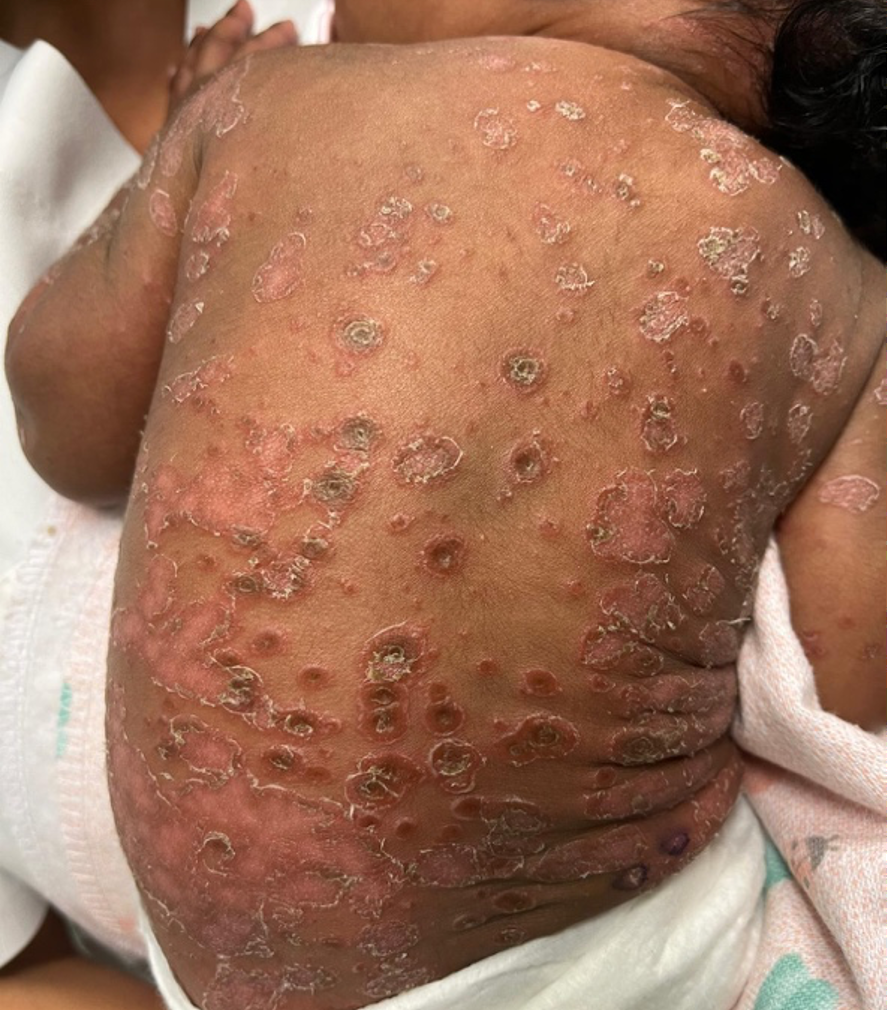
Five days later, she was readmitted with worsening lesions and irritability. At that time, she had scaling lesions of the face and trunk (Figure 1) and apparently abraded lesions in the neck folds, retroauricular fold, and umbilical area, with erythema and serous exudate, without associated fever. Intravenous flucloxacillin was reinstituted without meaningful improvement. Additional laboratory investigations, including immunoglobulin levels, were normal.
Due to the exuberant and atypical evolution, skin biopsy was performed to clarify the diagnosis. The infant was discharged home with oral flucloxacillin and topical hydrocortisone. She maintained regular outpatient follow-up with sustained improvement. Histopathology revealed dermatitis with a mixed reaction pattern (psoriasiform and spongiotic with neutrophils), suggesting, as differential diagnosis, impetiginised eczematous dermatitis, psoriasis or nutritional dermatitis. Zinc deficiency was then excluded. The lesions improved with regular application of an emollient and methylprednisolone aceponate as required, and the infant remained stable, without new lesions, for more than 6 months. Thus, TNPM with seborrhoeic eczema lesions and associated impetigo was assumed, and the infant was discharged from Dermatology follow-up.
Learning Points and Discussion: An exuberant, widespread and delayed presentation of TNPM can mimic other diagnoses — particularly when a second dermatosis (such as seborrhoeic eczema) is superimposed — prompting invasive investigation and avoidable antibiotic exposure. Skin biopsy is rarely necessary in TNPM but is useful when the course is atypical. Although TNPM is more frequent in neonates with darker skin, this case highlights the challenges in diagnosing common dermatological conditions in those with higher skin phototypes.