Background: Hirschsprung disease (HD) is a rare congenital disorder of the enteric nervous system, caused by impaired neural crest cell migration during intestinal development. This leads to aganglionosis of the distal bowel, most frequently affecting the rectosigmoid colon. Its most feared complication is the Hirschsprung-associated Enterocolitis (HAEC), the leading cause of mortality and morbidity.
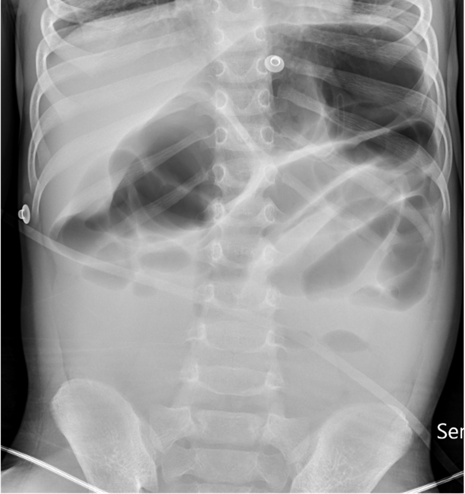
Case presentation: A 4-year-old boy, with a history of HD treated two years prior with a Swenson pull-through procedure, presented to the paediatric emergency department with a one-day history of abdominal pain, fetid diarrhoea, and vomiting. No other symptoms were recorded. Physical examination revealed no abnormalities. Oral antiemetics and hydration were administered. During hospitalization, he remained lethargic with progressive abdominal distension, for which intravenous fluids were initiated for maintenance and replacement of ongoing losses. Initial laboratory tests and blood cultures were unremarkable. Gastrointestinal molecular panel was positive for norovirus, with a negative stool culture. Abdominal radiography showed marked dilatation of bowel loops and faecal loading (Figure 1). Within six hours, he deteriorated rapidly with the onset of fever, tachycardia, signs of poor peripheral perfusion, and hypotension. Laboratory reassessment showed severe metabolic acidosis (pH and HCO3 below the measurable range), hyperlactatemia (9.8 mmol/L), acute kidney injury (creatinine 0.9 mg/dL; 2.25-fold increase from baseline), and elevated inflammatory markers (C-reactive protein 27.7 mg/dL and procalcitonin >100 ng/mL), findings consistent with severe HAEC presenting with mixed hypovolemic/septic shock. Resuscitation required a total of 60 mL/kg of crystalloid boluses, and ceftriaxone and metronidazole were started. Gastrointestinal decompression was performed via nasogastric tube placement, with drainage of faecal material, and rectal enema, with explosive discharge of stools. He was transferred to the paediatric intensive care unit. Further stabilization required three additional crystalloid boluses and aggressive fluid therapy (up to 3500 mL/m2/day), supplemented with calcium and potassium, without the need for vasoactive support. Antibiotics were adjusted to cefotaxime, metronidazole, and gentamicin. Serial rectal irrigations were performed, with abundant faecal output. Gastric residual progressively decreased, alongside marked improvement in abdominal distension. The patient was discharged after 12 days, clinically and analytically stable, with normalization of renal function.
Discussion: HAEC is a complication which may happen both before and after surgery, in 20-40% of HD patients. A high index of clinical suspicion is required for timely diagnosis and management. During the initial stages, clinical presentation is non-specific and can mimic acute gastroenteritis. However, patients may rapidly progress to mixed hypovolemic/distributive (septic) shock. Management relies on aggressive fluid therapy for haemodynamic stabilization and gastrointestinal source control. Broad-spectrum antibiotics are mandatory for seven to ten days, with coverage of anaerobes and enteric gram-negative organisms. Gastrointestinal decompression is crucial, with nasogastric tube placement, serial rectal irrigations, and bowel rest. If medical management is not sufficient, or in case of bowel perforation, peritonitis or pneumatosis, surgery might be needed. In this case, HAEC (Pastor/Delphi HAEC score of 11) occurred two years after surgery, likely triggered by norovirus infection.