Background: Lithobezoar is a rare gastrointestinal bezoar resulting from the accumulation of ingested stones. In children, it is most commonly associated with pica, which may occur in the setting of iron deficiency, autism spectrum disorder (ASD), developmental disorders, or underlying malabsorptive conditions such as celiac disease.
Objective: To describe the clinical presentation, underlying etiologies, radiological findings, and management of two pediatric patients with lithobezoar associated with pica.
Materials and Methods: We retrospectively reviewed the clinical presentation, laboratory investigations, imaging findings, underlying diagnoses, management, and follow-up of two pediatric patients diagnosed with lithobezoar at our center. Relevant clinical and laboratory data were collected from medical records.
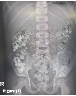
Results: Two boys, aged 8 and 10 years, were identified. The first patient presented with recurrent abdominal pain, constipation, pica, poor growth, and severe microcytic iron deficiency anemia. Abdominal radiography demonstrated multiple radiopaque materials within the colon without evidence of obstruction as shown in figure (1). Further evaluation revealed strongly positive anti-tissue transglutaminase IgA and anti-endomysial antibodies, confirming celiac disease. He was treated with a gluten-free diet, intravenous iron, and laxatives, resulting in clinical improvement, normalization of iron stores, and complete radiographic resolution.
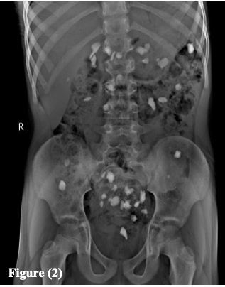
The second patient was a 10-year-old boy with ASD who had ingested more than 20 small stones. He was asymptomatic, but abdominal radiography demonstrated multiple radiopaque foreign bodies throughout the small and large intestines as shown in figure (2). A three-year history of pica and selective eating behavior was identified. He was managed conservatively with laxatives, behavioral monitoring, and developmental pediatric follow-up. Neither patient required endoscopic or surgical intervention.
Conclusion: Pediatric lithobezoar is rare but should be considered in children with pica and characteristic radiographic findings. Pica may be associated with different underlying conditions, including iron deficiency secondary to celiac disease and ASD. Children presenting with pica, particularly those with unexplained iron deficiency anemia or poor growth, should undergo appropriate evaluation for underlying disorders. Conservative management may be successful in selected asymptomatic patients without intestinal obstruction.