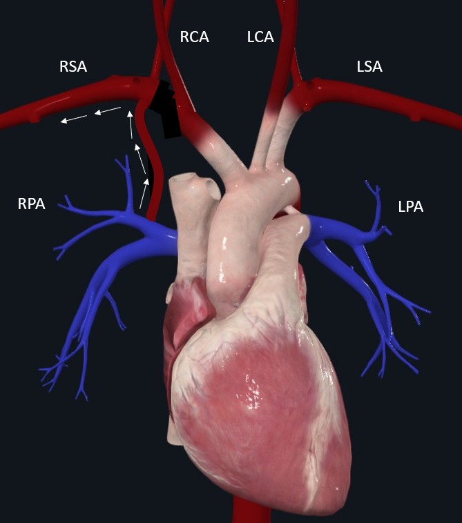
Background: DiGeorge syndrome (DGS) is a genetic disorder caused by a hemizygous 22q11 microdeletion, with a wide variability of phenotypic features. The clinical presentation is well known with facial dysmorphism, thymic hypoplasia/aplasia, immune deficiency, hypoparathyroidism and congenital heart disease (CHD). DGS is the second leading chromosomal cause of CHD after trisomy 21. The most common CHD includes conotruncal defects (such as tetralogy of Fallot). Aortic arch anomalies (AAA) have been described in DGS, including cervical aortic arch, double aortic arch, right-sided aortic arch, and aberrant right or left subclavian artery. But we report a rare case of an isolated right subclavian artery (IRSA) arising from the right pulmonary artery via a right-sided ductus arteriosus; therefore, the right arm was perfused by poorly oxygenated blood from the pulmonary artery and was underdeveloped in comparison to the left arm. The patient showed a subclavian and vertebral artery steal. The patient underwent successful surgical repair. Literature on this IRSA condition is scarce, especially for isolated defects.
Case presentation summary: An asymptomatic newborn (gestational age 39 weeks) was screened by pre and post-ductal oxygen saturation (pre-SpO2 85%, post-SpO2 99%). Apgar scores were 9/10 (1/ 5 minutes). Clinical examination, fetal and neonatal echocardiography were, apparently, normal. In the first few months of life, underdevelopment of the right arm was observed. An ECG-gated contrast-enhanced CT was performed and showed isolation of the right subclavian artery, which arises via a right-sided ductus from the right pulmonary artery (Figure 1). This anomaly caused subclavian steal and poorly oxygenated perfusion of the arm. No thymus was observed on the CT scan. Genetic studies confirmed a 22q11 deletion. No hypocalcaemia or immunodeficiency was detected. Our patient underwent a successful surgical repair (17 months old), the right subclavian artery was reimplanted to the aortic arch. The surgery confirmed the absence of the thymus. The patient is being monitored by cardiologists, endocrinologists, and immunologists.
Learning points discussion:
1.- Aortic arch anomalies have been described in DiGeorge syndrome, and almost always appear with coexisting congenital heart disease, most often Tetralogy of Fallot.
2.- The IRSA arising from the right pulmonary artery, without coexisting structural heart defects, is the rarest aortic arch anomaly.
3.- Although IRSA can be asymptomatic, these patients can present with vertebrobasilar insufficiency (syncopal episodes), limb deformities, or pulmonary overcirculation.
4.- The IRSA has a successful surgical repair, although there are no surgical guidelines for this condition